
|
Fig 4
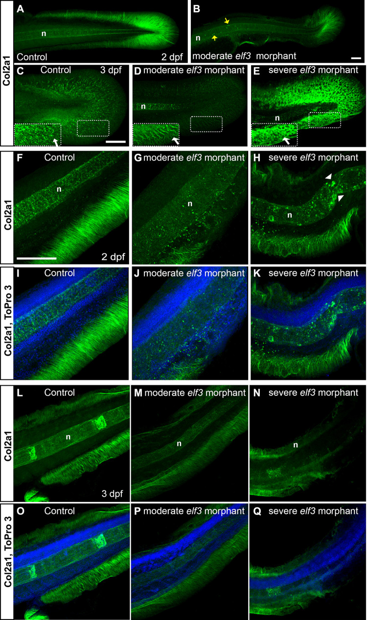
(A-E) 3D reconstruction of the confocal images of the Col2?1 antibody stained embryos show dense Col2?1 fibrils radially distributed throughout the whole caudal fin in the control (A, C) and less dense, irregularly distributed fibrils in moderately affected morphants (B, D); severely affected morphant showed abnormal accumulation of Col2?1, but no fibrillary pattern (E). Insets show high magnification of the marked rectangle area. yellow arrow: abnormal Col2?1 accumulation within notochord; white arrow: Col2?1 fibrils. (F-Q) Optical section of the confocal images of the Col2?1 antibody stained embryos show severely disrupted and disorganization median fin fold collagen fibrils in the elf3 morphants at 2 dpf (G, H, J, K) and 3 dpf larva (M, N, P, Q) and nicely organized, thick fibrils in the control (F, I, L, O). Scale bar for A, B = 50 ?m; for C-E = 100 ?m; and for F-Q = 100 ?m.