|
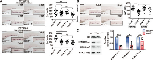
Inhibition of PRC1 components is associated with disruption of neutrophils development.A WISH for lyz (3 dpf) after UNC3866 treatment showing lyz+ cells decreased after 50 μM UNC3866 treatment. Quantification of lyz+ cells in asxl1+/+ and asxl1−/− tails (scale bar, 200 μm; black boxes show enlarged images; asxl1+/+, n = 16, asxl1−/−, n = 18, asxl1+/+ T, n = 20, asxl1−/− T, n = 15; one-way ANOVA followed by LSD Fisher’s post hoc test, *p < 0.05, **p < 0.01, ***p < 0.001; error bars, mean ± SD). T treated, ns not significant. WISH of lyz (3 dpf) after 10 μM PRT4165 treatment showing lyz+ cells decreased after PRT4165 treatment. Quantification of lyz+ cells in asxl1+/+ and asxl1−/− tails (scale bar, 200 μm; black boxes show enlarged images; asxl1+/+, n = 12, asxl1−/−, n = 15, asxl1+/+ T, n = 10, asxl1−/− T, n = 14; one-way ANOVA followed by LSD Fisher’s post hoc test, *p < 0.05, **p < 0.01, ***p < 0.001; error bars, mean ± SD). T treated, ns not significant. B WISH for lyz (3 dpf) after bmi1a mRNA injection showing reduced lyz expression was rescued by bmi1a mRNA injection. Quantification of lyz+ cells in asxl1+/+ and asxl1−/− tails (about 0.1 ng mRNA/embryo; scale bar, 200 μm; black boxes show enlarged images; control: asxl1+/+, n = 12, asxl1−/−, n = 18; bmi1a: asxl1+/+, n = 18, asxl1−/−, n = 10; one-way ANOVA followed by LSD Fisher’s post hoc test, *p < 0.05, **p < 0.01, ***p < 0.001; error bars, mean ± SD; T treated, ns not significant). C Western blot of H2AK119ub, H3K4me3 and H3K27me3 in 2 dpf asxl1+/+ and asxl1−/− embryos and quantification of Western blot data (input embryos: about ten embryos/well, performed with three replicates; two-tailed Student’s t test, ns not significant; error bars, mean ± SD, internal control, H3).
|