
Figure 2
- ID
- ZDB-FIG-210319-74
- Publication
- Lai et al., 2021 - MicroRNA-21 Plays Multiple Oncometabolic Roles in the Process of NAFLD-Related Hepatocellular Carcinoma via PI3K/AKT, TGF-β, and STAT3 Signaling
- Other Figures
- All Figure Page
- Back to All Figure Page
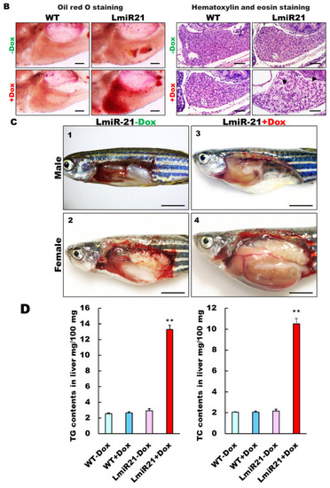
Characterization of hepatic steatosis in LmiR21 larvae and adult. (A) Whole-mount oil-red O (ORO) staining of WT � Dox and LmiR21 � Dox larvae at 21 dpf. Lipid contents in livers and swim bladders were stained by ORO (up). Scale bar: 2 mm. Percentages of WT � Dox and LmiR21 � Dox larvae with weak, moderate and strong levels of hepatic steatosis at 21 dpf (down). Scale bar: 200 ?m. (B) Representative ORO images of liver in 21 dpf larvae (left). Scale bar: 500 ?m. Hematoxylin-eosin (H&E) staining of livers at 21 dpf LmiR21 + Dox larvae show abnormally shaped hepatocytes with lipid vacuoles (arrows) compared with that in the controls at the same stage. Scale bar: 100 ?m. (right). (C) Gross liver phenotypes at 4 months post fertilization (mpf) LmiR21 � Dox. Normal hepatic morphology is clearly composed of lobes in LmiR21 ? Dox adult (panels 1,2). Liver hyperplasia with yellow and greasy phenotypes in LmiR21 + Dox adult liver (panels 3,4). Scale bar: 5 mm. (D) Triglyceride (TG) and total cholesterol (TC) in the livers is greater in LmiR21 + Dox than in the controls (n = 3). Statistically significant differences from LmiR21 ? Dox were denoted by ** (p < 0.01). (E) H&E staining of liver tissue from WT � Dox and LmiR21 � Dox at 4 mpf depicting morphological changes resulting from accumulation of microvesicular steatosis. Scale bar: 50 ?m. (left). ORO staining of liver cryosections from WT � Dox and LmiR21 � Dox adults at 4 mpf. Scale bar: 50 ?m. An abundance of lipid accumulation is observed in LmiR21 + Dox compared to that in controls. (right). (F) Representative liver sections showing ballooned hepatocytes (large arrow) and lipid vacuoles (star) with Masson?s trichrome (left), and Picrosirius red (right). Scale bar: 50 ?m. |