|
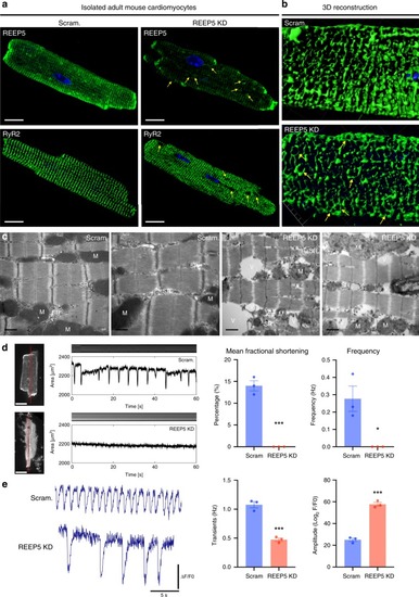
In vitro REEP5 depletion leads to SR vacuolization and sarcomeric dysfunction in adult mouse cardiac myocytes.a Immunofluorescence of adult mouse cardiac myocytes stained with REEP5 and RyR2 48 h post viral transduction with REEP5 shRNA. Scale, 20 μm. b Three-dimensional reconstruction analysis of RyR2 staining of the SR in Scram and REEP5-depleted myocytes. c Transmission electron microscopy in REEP5-depleted adult mouse cardiac myocytes revealed SR vacuoles and disrupted SR membranes compared to scram controls. M mitochondria, SR sarcoplasmic reticulum, TT T-tubule, V vacuoles. Scale, 0.5 nm. d Optical measurements of spontaneous myocyte contractility in scram and REEP5-depleted adult mouse cardiac myocytes revealed a significant decrease in both fractional shortening measurements and frequency in REEP5-depleted myocytes. Left: still images of representative cardiac myocytes. Scale, 20 μm. Red line indicates region of image used to generate kymographs shown above contractile pulses tracings. n = 30 cells examined over three independent experiments; data are presented as mean ± SEM. e Ca2+ imaging of myocytes showing the frequency of Ca2+ waves and Ca2+ transients amplitude 48 h post viral transduction with REEP5 shRNA, n = 40–50 cells examined per condition over 3 independent experiments. Asterisks indicate a statistically significant p value in a Tukey’s multiple comparison analysis where *p < 0.05 and ***p < 0.001; data are presented as mean ± SEM. All images shown are representative of approximately 30–40 total images captured per condition.
|