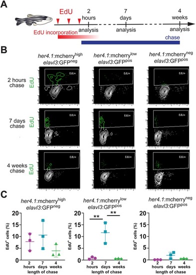
Newborn neurons retain the radial glia-derived reporter signal transiently. (A) Scheme of experimental design, indicating the timing of EdU injections (red arrowheads), EdU incorporation (red bar), EdU chase (blue bar) and analysis timepoints after the last EdU injection. (B) Flow cytometry plots showing EdU (green) incorporation in sorted her4.1:mCherryhigh/elavl3:GFPneg cells (left), her4.1:mCherrylow/elavl3:GFPpos cells (middle) and her4.1:mCherryneg/elavl3:GFPpos cells (right) after 2 h (top), 7 days (middle) or 4 weeks (bottom) of chase. There is robust EdU labeling in her4.1:mCherrylow/elavl3:GFPpos after 7 days, but not after 2 h or 4 weeks chase. (C) Quantification of EdU labeling her4.1:mCherryhigh/elavl3:GFPneg cells (left), her4.1:mCherrylow/elavl3:GFPpos cells (middle) and her4.1:mCherryneg/elavl3:GFPpos cells (right) after 2 h (magenta), 7 days (blue) or 4 weeks (green). Single data points are shown; n=3; data are mean±s.e.m. **P<0.01 by one-way ANOVA and Bonferroni's post hoc test.
|
