
FIGURE
Fig. S3
- ID
- ZDB-FIG-190814-24
- Publication
- Reuter et al., 2019 - Fgf3 is crucial for the generation of monoaminergic cerebrospinal fluid contacting cells in zebrafish
- Other Figures
- All Figure Page
- Back to All Figure Page
Fig. S3
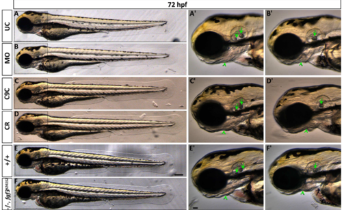
Live images showing morphology of 72 hpf embryos after fgf3 impairment with characteristic ear and craniofacial malformations. (A,B) Uninjected control (UC) and fgf3 morpholino injected (MO) siblings. (C,D) Cas9 injected control (C9C) and fgf3 CRISPR/Cas9 injected (CR) siblings. (E,F) Wildtype (+/+) and homozygous fgf3t24152 mutant (-/-) siblings. Boxes indicate magnified area shown in A’-F’. After fgf3 impairment the two otoliths fuse (arrow) and the lower jaw bones are malformed (arrow heads). Lateral views, anterior to the left. Scale bar in E, 100 μm; in E’, 50 μm. |
Expression Data
Expression Detail
Antibody Labeling
Phenotype Data
| Fish: | |
|---|---|
| Knockdown Reagents: | |
| Observed In: | |
| Stage: | Protruding-mouth |
Phenotype Detail
Acknowledgments
This image is the copyrighted work of the attributed author or publisher, and
ZFIN has permission only to display this image to its users.
Additional permissions should be obtained from the applicable author or publisher of the image.
Full text @ Biol. Open