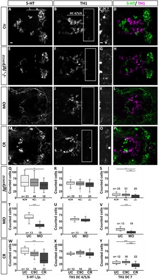
Quantification of the number of serotonergic cells in the intermediate (i.)/posterior (p.) clusters and of dopaminergic cells in the DC 4/5/6 and DC 7 clusters in the hypothalamus at 72 hpf after fgf3 impairment. (A–P) Confocal maximum intensity projections from wild-type controls (Ctr), homozygous fgf3t24152 mutants (−/−), fgf3 morphants (MO) or fgf3 CRISPR/Cas9-injected embryos (CR) immunostained for 5-HT (green) and TH1 (magenta) shown as single channels and merged. C,G,K and O show boxed areas in B,F,J and N, respectively, with adjusted brightness and contrast to reveal faint TH1 immunoreactive cells of the DC 7 cluster. Ventral views, anterior to the left. Scale bars: 10 µm. (Q–Y) Quantifications of 5-HT and TH1 positive cells after fgf3 impairment and in control siblings. The number of serotonergic cells was counted in the i./p. clusters as indicated by the line in A. The number of dopaminergic cells was counted in the DC 4/5/6 and DC 7 clusters as indicated by the lines in B and C. Tukey boxplots show median, 25–75% percentile, IQR whiskers and outliers. n=number of analysed individuals. +/−, heterozygous fgf3t24152 mutants; UC, uninjected siblings; C9C, injected with Cas9 only. *P>0.05, **P>0.01, ***P>0.001.
|
