
Fig. S5
- ID
- ZDB-FIG-190812-40
- Publication
- Chen et al., 2019 - Cerebrovascular Injuries Induce Lymphatic Invasion into Brain Parenchyma to Guide Vascular Regeneration in Zebrafish
- Other Figures
- All Figure Page
- Back to All Figure Page
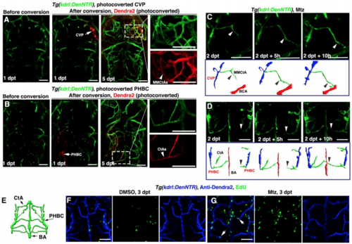
The regenerating blood vessels arise from residual blood vessels through cell proliferation and migration, Related to Figure 1. (A and B) At 1 dpt after Mtz treatment, the residual blood vessels CVP (A, arrow) and PHBC (B, arrow) were photoconverted from green to red fluorescence. At 5 dpt, retentions of the Dendra2-red fluorescence were detected in nascent blood vessels MMCtAs (A, arrowhead, n=9/10) and CtAs (B, arrowhead, n=8/10), respectively. (C and D) Live imaging of the nascent MMCtA and CtA formation. Nascent MMCtA was observed to migrate from CVP to BCA (C, arrowheads, n=10/10), while nascent CtA migrated from PHBC to BA (D, arrowheads, n=10/10), accompanied by explanatory schematic diagrams. (E–G) Illustration of hindbrain vascular network indicate the image area in following panels (E). In contrast to DMSO treatment in which brain BECs rarely proliferated (F, n=20/20), many of nascent BECs were positive for EdU labeling at 3 dpt after Mtz treatment (G, arrows, n=19/21). All images are dorsal view, anterior upward. Scale bar, 50 μm. BA, basilar artery; BCA, basal communicating artery; CtA, central artery; CVP, choroidal vascular plexus; MMCtA, middle mesencephalic central artery; PHBC, primordial hindbrain channel. |
Reprinted from Developmental Cell, 49(5), Chen, J., He, J., Ni, R., Yang, Q., Zhang, Y., Luo, L., Cerebrovascular Injuries Induce Lymphatic Invasion into Brain Parenchyma to Guide Vascular Regeneration in Zebrafish, 697-710.e5, Copyright (2019) with permission from Elsevier. Full text @ Dev. Cell