
Fig. 1
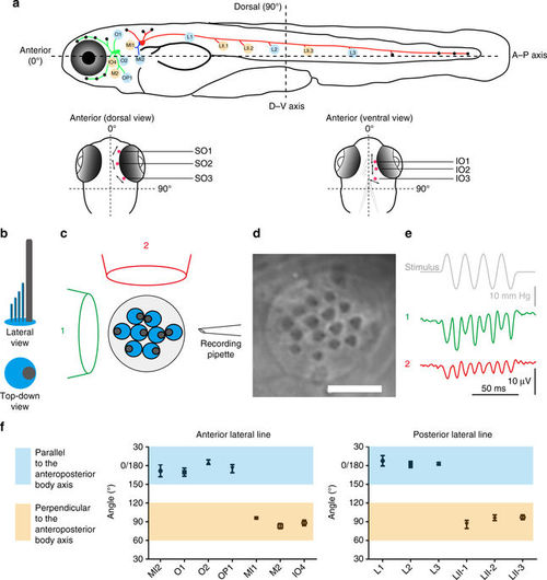
Axes of best sensitivity for anterior and posterior lateral line neuromasts. a Schematic of the zebrafish lateral line at ~6?dpf. (top) Side view. Neuromasts that have been scored are blue or tan and correspond to the graph below. ALL(green), PLL(red), and medial?lateral-line (blue) ganglia contact neuromasts. (bottom) Dorsal and ventral views of the head. Supraorbital (SO) neuromasts 1 to 3 and infraorbital (IO) neuromasts 1 to 3 are marked in red. The mean angle of the orientation of the axis of best sensitivity relative to the A?P body axis of each neuromast is presented as a double-headed arrow. b?e Evaluation of the axis of best sensitivity for a neuromast. b Schematics of hair bundles. Gray and blue protrusions are a kinocilium and stereocilia, respectively. c Experimental configuration: a top-down view of a neuromast with a fluid jet pipette that provides stimuli (1) parallel to the axis along which the bevel-shaped hair bundles face opposite directions or (2) orthogonal to that axis. The pipettes in the diagram are not to scale. d DIC image of hair bundles of a posterior neuromast from which stimulus-evoked microphonic potentials were recorded. Scale bar?=?5??m. e Recordings from an A?P oriented L3 neuromast with fluid jet pipettes stimulating, serially, from two different directions depicted in c. f Graphs of mean angle of the orientation of the axis of best sensitivity relative to the A?P body axis of each anterior and posterior neuromast position?�?SEM (n? =?4?7). Each axis with a range from 60� to 120� from the A?P body axis is defined perpendicular (tan). Each axis with a range from 0� to 30� or 150� to 180� from the A?P body axis is defined as parallel (blue). ALL nomenclature: opercular (OP), mandibular (M), infraorbital (IO), supraorbital (SO), middle (MI) and otic (O). PLL nomenclature: L neuromasts are A?P oriented; LII neuromasts are D?V oriented. These different neuromast types are derived from different migrating primordia during development |