
Fig. 6
- ID
- ZDB-FIG-180302-18
- Publication
- Seritrakul et al., 2017 - Tet-mediated DNA hydroxymethylation regulates retinal neurogenesis by modulating cell-extrinsic signaling pathways
- Other Figures
- All Figure Page
- Back to All Figure Page
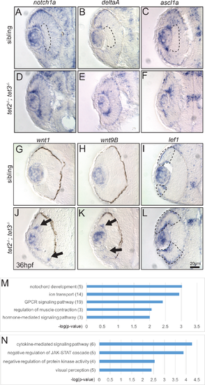
Fig 6. Gene expression is altered in tet2-/-;tet3-/- mutants at 36hpf and differentially expressed genes include those encoding components of the Notch and Wnt pathways. (A-C) In sibling embryos at 36hpf, transcripts encoding components of Notch pathway (notch1a, deltaA, and ascl1a) are expressed in the eye but excluded from the inner part of central retina where cells have exited the cell cycle and differentiated (dotted area). (D-F) In tet2-/-;tet3-/- mutants, these genes are expressed throughout the retina without a clear ?zone? of differentiation (n>8). (G,H,J,K) The expression domains of wnt1 and wnt9B, are expanded in tet2-/-;tet3-/- mutants (arrows; n = 7 for wnt1, n = 8 for wnt9b), consistent with RNA-Seq data. (I,L) Similarly, lef1, a downstream readout of Wnt pathway activity, is normally expressed in the peripheral edge of the retina, and this zone of expression is expanded in tet2-/-;tet3-/- mutants (dotted areas; n>8). (M-N) Gene ontology (GO) analysis for biological pathways was performed using DAVID. Numbers in parentheses indicate number of genes enriched in each pathway. P-value cutoff = 0.01. |
| Genes: | |
|---|---|
| Fish: | |
| Anatomical Terms: | |
| Stage: | Prim-25 |
| Fish: | |
|---|---|
| Observed In: | |
| Stage: | Prim-25 |