Fig. 1
- ID
- ZDB-FIG-170424-4
- Publication
- Schmitner et al., 2017 - ptf1a+ , ela3l- cells are developmentally maintained progenitors for exocrine regeneration following extreme loss of acinar cells in zebrafish larvae.
- Other Figures
- All Figure Page
- Back to All Figure Page
|
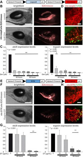
Exocrine-tissue-specific zebrafish cell ablation systems. (A) Construct used to generate ela:casp8 transgenics (black arrowheads indicate promoter direction). (B) Brightfield and epifluorescence images of 7?dpf larvae showing E2Crimson signals in untreated control animals and after ablation with 5?µM Dim for 48?h. (C) Relative expression levels of ela3l (left) and trypsin (right) mRNA as revealed by qPCR analyses of whole-embryo RNA preparations of control larvae and larvae treated with 1.6, 5 and 8?µM Dim for 48 and 96?h, normalized to ?-actin levels. (D) Confocal projection of untreated 5?dpf larva and larva treated for 8?h with 5?µM Dim labelled by TUNEL staining (green). (E) Construct used to generate the ela:DTR transgenics. (F) Brightfield and epifluorescence images of 9?dpf untreated larvae showing exocrine-specific expression of E2Crimson and strongly reduced expression of E2Crimson after incubation in 15?µg/ml DT for 96?h. (G) Relative expression levels of ela3l (left) and trypsin (right) mRNA as revealed by qPCR analyses of whole-embryo RNA preparations of control larvae and larvae treated with 10 and 15?µg/ml DT for 48 and 96?h, normalized to ?-actin levels. (H) Confocal projection of untreated 7?dpf larva and larva treated for 72?h with 15?µg/ml DT labeled by TUNEL staining (green). Values are mean+s.e.m. of n=5 embryos per sample and >3 biological replicates. Scale bars: 150?µm (B,F) and 20?µm (D,H). |

