
Fig. S2
- ID
- ZDB-FIG-170424-12
- Publication
- Schmitner et al., 2017 - ptf1a+ , ela3l- cells are developmentally maintained progenitors for exocrine regeneration following extreme loss of acinar cells in zebrafish larvae.
- Other Figures
- All Figure Page
- Back to All Figure Page
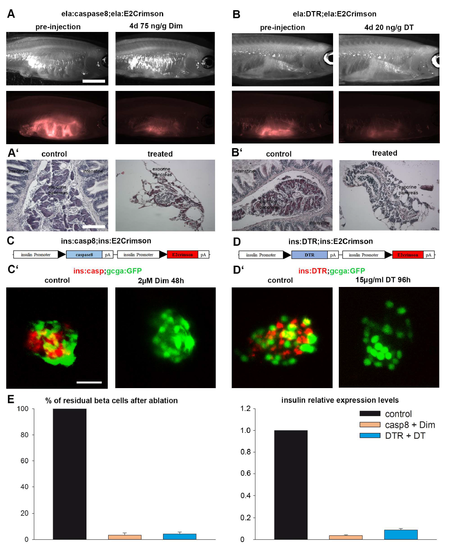
Ablation of exocrine tissue in adult zebrafish and beta cells in embryos. A: brightfield and epifluorescence images of ela:casp8 adults before intraperitoneal injection of 75ng/g Dim and 4 days after injection showing the loss of E2crimson fluorescence. A': histological sections of an intact exocrine pancreas of an untreated fish and of a disrupted exocrine pancreas 4 days after treatment. B: brightfield and epifluorescence images of ela:DTR adults before intraperitoneal injection of 20 ng/g DT and 4 days after injection showing the loss of E2crimson fluorescence. B': histological sections of an intact exocrine pancreas of an untreated fish and of an ablated exocrine pancreas four days after treatment. C: construct to target expression to the beta cells and C' confocal projections of endocrine islets of control and treated tg(ins:casp8;gcga:eGFP) embryos at 6 dpf; D: construct to establish the ins:DTR transgenics and D' confocal projections of endocrine islets of control and treated tg(ins:DTR;gcga;eGFP) embryos at 8 dpf. E: relative amount of beta cells and relative expression levels of insulin of control and [sic]. |