
Fig. 7
- ID
- ZDB-FIG-170424-10
- Publication
- Schmitner et al., 2017 - ptf1a+ , ela3l- cells are developmentally maintained progenitors for exocrine regeneration following extreme loss of acinar cells in zebrafish larvae.
- Other Figures
- All Figure Page
- Back to All Figure Page
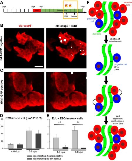
Proliferation of exocrine cells during regeneration is dependent on Wnt signaling. Confocal projections of Tg(ela:casp8) crossed to Tg(hsp70l:dkk1-eGFP) and treated with 5?�M Dim. During regeneration (4-6?dpa or 6-8?dpa) larvae were heat shocked twice to induce dkk1 and they were additionally injected with EdU. (A) Timeline indicating treatments and heat shock (flames). (B) Control littermate negative for dkk1-eGFP. (C) Larvae expressing dkk1-eGFP showed reduced numbers of EdU+ cells. Volume of E2Crimson cells (D) and EdU+ E2Crimson+ cells (E) in larvae expressing dkk1-eGFP and controls at 6 and 8?dpa during regeneration, showing that the induced expression of dkk1 reduces the number of EdU+ exocrine cells (N>4 larva for each time point). Mean+ s.e.m.; **P<0.001. (F) Model of exocrine cell regeneration after complete removal of exocrine cells. Scale bar: 20?�m. |