
Fig. 1
- ID
- ZDB-FIG-170104-8
- Publication
- Jiménez-Amilburu et al., 2016 - In Vivo Visualization of Cardiomyocyte Apicobasal Polarity Reveals Epithelial to Mesenchymal-like Transition during Cardiac Trabeculation
- Other Figures
- All Figure Page
- Back to All Figure Page
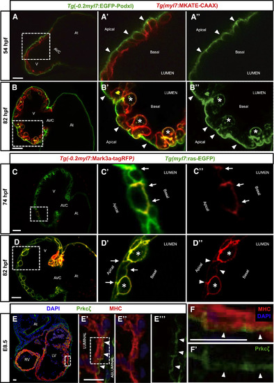
Establishment of Apicobasal Polarity in CMs (A?B'') Confocal images (mid-sagittal sections) of Tg(?0.2myl7:EGFP-podxl);Tg(myl7:MKATE-CAAX) zebrafish hearts at 54 (A?A'') and 82 (B?B'') hpf. Boxed area in (A) is shown in (A') and (A''). EGFP-Podxl is restricted to the abluminal membrane of CMs (A' and A'', arrowheads). Boxed area in (B) is shown in (B') and (B''). EGFP-Podxl is observed all around the membrane of delaminated CMs (B' and B'', asterisks), while it remains apical in compact-layer CMs (B' and B'', arrowheads). (C?D'') Confocal images (mid-sagittal sections) of 74-hpf (C?C'') and 82-hpf (D?D'') Tg(myl7:ras-EGFP) larvae injected with ?0.2myl7:mark3a-TagRFP-T plasmid. Boxed area in (C) is shown in (C') and (C''). Mark3a-TagRFP-T, a basolateral marker, is restricted to the luminal and lateral membranes of CMs (C and C'', arrows). Boxed area in (D) is shown in (D') and (D''). Mark3a-TagRFP-T is observed all around the membrane of delaminated CMs (D' and D'', asterisks), while it remains basolateral in compact-layer CMs (D' and D'', arrowheads). (E?E'') E8.5 mouse heart co-stained for Prkc? (green), myosin heavy chain (MHC) (red), and DAPI (blue). High-magnification images show the localization of the apical protein Prkc? in the abluminal membrane of CMs (E' and E''', arrowheads). (F?F') High-magnification images of a single mouse cardiomyocyte showing abluminal localization of Prkc? (F and F', arrowheads). Boxed area in (E') is shown in (F) and (F'). At, atrium; V, ventricle; AVC, atrioventricular canal; RV, right ventricle; LF, left ventricle. Scale bars, 20 ?m. |
| Genes: | |
|---|---|
| Fish: | |
| Anatomical Terms: | |
| Stage Range: | Long-pec to Protruding-mouth |