
Fig. S5
- ID
- ZDB-FIG-170104-19
- Publication
- Jiménez-Amilburu et al., 2016 - In Vivo Visualization of Cardiomyocyte Apicobasal Polarity Reveals Epithelial to Mesenchymal-like Transition during Cardiac Trabeculation
- Other Figures
- All Figure Page
- Back to All Figure Page
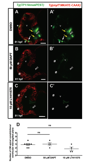
Notch inhibition does not affect cardiomyocyte apical constriction (A-C') Confocal images (mid-sagittal sections) of 81 hpf Tg(myl7:MKATE-CAAX);Tg(TP1:VenusPEST) zebrafish hearts. (A-A') DMSO-treated larvae showing TP1:VenusPEST positive cardiomyocytes (A and A', asterisks), endocardial cells (A and A', white arrows) and AV valve cells (A and A', yellow arrows). (B- C'') Embryos were treated starting at 48 hpf with 50 ?M DAPT (B-B') or 10 ?M LY411575 (C-C') and imaged at 81 hpf. In both sets of experimental animals, Notch activation was not observed in cardiomyocytes (B' and C', asterisks). (D) Number of apical constrictions after 50 ?M DAPT or 10 ?M LY411575 inhibitor treatments at 79 hpf. Each dot represents one heart. Data are shown as mean ± SEM. ns, no significant differences by Student's t-test. V, ventricle; AVC, atrioventricular canal. Scale bars, 20 ?m. |