
|
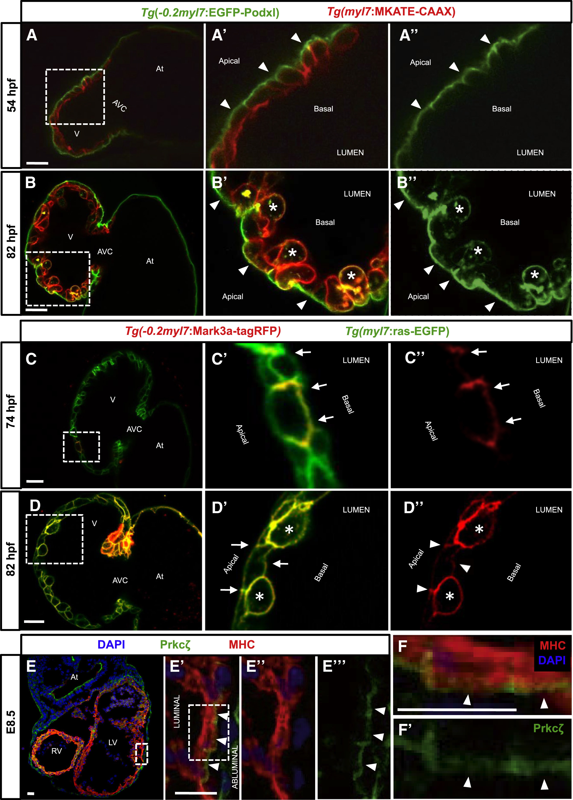
Fig. 1
Establishment of Apicobasal Polarity in CMs
(A?B'') Confocal images (mid-sagittal sections) of Tg(?0.2myl7:EGFP-podxl);Tg(myl7:MKATE-CAAX) zebrafish hearts at 54 (A?A'') and 82 (B?B'') hpf. Boxed area in (A) is shown in (A') and (A''). EGFP-Podxl is restricted to the abluminal membrane of CMs (A' and A'', arrowheads). Boxed area in (B) is shown in (B') and (B''). EGFP-Podxl is observed all around the membrane of delaminated CMs (B' and B'', asterisks), while it remains apical in compact-layer CMs (B' and B'', arrowheads).
(C?D'') Confocal images (mid-sagittal sections) of 74-hpf (C?C'') and 82-hpf (D?D'') Tg(myl7:ras-EGFP) larvae injected with ?0.2myl7:mark3a-TagRFP-T plasmid. Boxed area in (C) is shown in (C') and (C''). Mark3a-TagRFP-T, a basolateral marker, is restricted to the luminal and lateral membranes of CMs (C and C'', arrows). Boxed area in (D) is shown in (D') and (D''). Mark3a-TagRFP-T is observed all around the membrane of delaminated CMs (D' and D'', asterisks), while it remains basolateral in compact-layer CMs (D' and D'', arrowheads).
(E?E'') E8.5 mouse heart co-stained for Prkc? (green), myosin heavy chain (MHC) (red), and DAPI (blue). High-magnification images show the localization of the apical protein Prkc? in the abluminal membrane of CMs (E' and E''', arrowheads).
(F?F') High-magnification images of a single mouse cardiomyocyte showing abluminal localization of Prkc? (F and F', arrowheads). Boxed area in (E') is shown in (F) and (F').
At, atrium; V, ventricle; AVC, atrioventricular canal; RV, right ventricle; LF, left ventricle. Scale bars, 20 ?m.