
Fig. 5
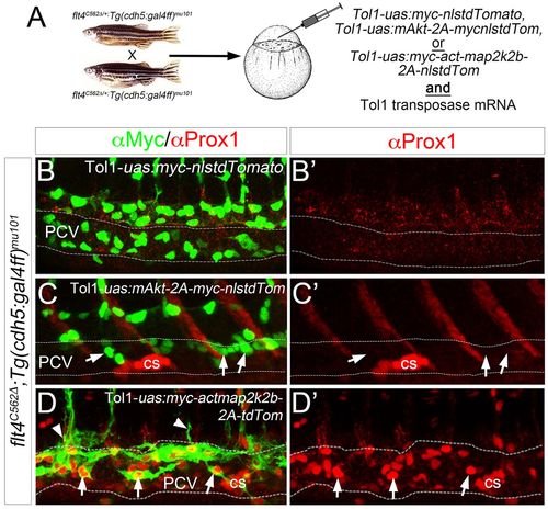
ERK activation rescues early lymphatic morphogenesis and differentiation in the absence of Flt4 signaling. (A) Schematic depicting injection strategy for rescue experiments. (B-D?) Confocal microscopy of Tg(cdh5:gal4ff)mu101;flt4C562? mutant embryos injected with Tol1-uas:myc-nlstdTomato (B), Tol1-uas:mAkt-2a-myc-nlstdTomato (C) or Tol1-uas:myc-actmap2k2b-2a-tdTomato (D) along with mRNA encoding Tol1 Transposase. (B-D) Overlay of Prox1 (red) and Myc (green) immunostaining to visualize transgene expression in injected embryos at 38?hpf. Arrows indicate transgene-expressing cells; arrowheads indicate transgene-expressing cells that are sprouting from the PCV. (B?-D?) Prox1 immunostaining (red) in same injected embryos as in B-D. Corpuscle of Stannius (cs) is indicated. Dashed lines outline the PCV. |