
Fig. S13
- ID
- ZDB-FIG-160304-38
- Publication
- Ye et al., 2016 - An insulin signaling feedback loop regulates pancreas progenitor cell differentiation during islet development and regeneration
- Other Figures
- All Figure Page
- Back to All Figure Page
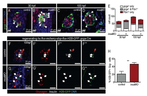
insulin knockdown increases α cell plasticity. (A-D) Confocal planes show control (A,C) and insaMO-injected (B,D) Tg(gcga:GFP) islets at 30 hpf (A,B) and 100 hpf (C,D) immunostained for Pdx1 (red) and DNA (blue). An increased number of double positive Gcg+ Pdx1+ cells are indicated by arrows. (E) Quantification of gcga:GFP+ (white) , gcga:GFP+ Pdx1+ (gray), and Pdx1+ (red) cells in control and insaMO-injected embryos at 30 hpf and 100 hpf. n≥7 for all time points. (F-G′′′) Confocal planes showing β cell regeneration after MTZ treatment in control (F-F′′′) and insaMO-injected (G-G′′) triply transgenic Tg(ins:Flag-NTR); Tg(hs:loxp-mcherry-loxp-H2BGFP); Tg(gcga:cre)s962 islets immunostained for Insulin (white) , GFP (green), Glucagon (red), and DNA (blue). Red arrows indicate H2B-GFP+ Glucagon+ cells, which have retained α cell character, while white arrows indicate H2B-GFP+ Glucagon- cells, which have lost α cell character during β cell regeneration. (H) Percentage of the H2B-GFPpositive cell population that is Gcg-negative in control (n=7) and insaMO-injected regenerating (n=13) islets. More lineage-labeled α cells lost glucagon expression in insaMO-injected islets than in control, suggesting increased plasticity. Student?s t-test was used in H to determine significance. |
Reprinted from Developmental Biology, 409(2), Ye, L., Robertson, M.A., Mastracci, T.L., Anderson, R.M., An insulin signaling feedback loop regulates pancreas progenitor cell differentiation during islet development and regeneration, 354-69, Copyright (2016) with permission from Elsevier. Full text @ Dev. Biol.