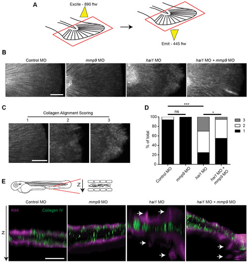
SHG imaging shows altered collagen alignment in hai1 morphants that is partially rescued by mmp9 depletion. (A) SHG imaging of type I/III collagen. Caudal fins were excited with an 890′w laser and collagen was detected at 445′w. (B) Representative z-projected images of stitched multiple ROI SHG z-stacks. The hai1 morphants display irregular collagen alignment. (C) In order to quantify alignment, SHG images illustrate scoring scheme, ranking severity of collagen mis-alignment for analysis from 1 to 3. (D) The hai1 morphants display a significant decrease in collagen alignment that could be partially rescued upon knockdown of mmp9 expression (MO1). (E) IHC of type IV collagen in the transgenic zebrafish Tg(krt4:tdTom). Epithelial extrusions were observed (arrows) in the hai1 morphants. Knockdown of mmp9 resulted in fewer observed extrusions. *P<0.05 and ***P<0.001. Scale bars: 50�m in B,C; 20�m in E. D represents the data from experiments performed in quadruplicate and scored by an individual, single-blind analyzer. D represents pooling with experimental numbers for Control MO=18, mmp9 MO=18, hai1 MO=20, hai1 MO+mmp9 MO=20.
|
