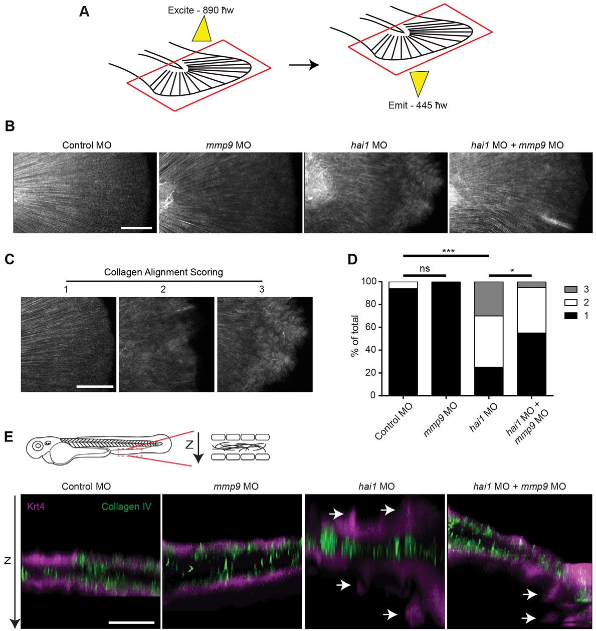
Fig. 3 SHG imaging shows altered collagen alignment in hai1 morphants that is partially rescued by mmp9 depletion. (A) SHG imaging of type I/III collagen. Caudal fins were excited with an 890′w laser and collagen was detected at 445′w. (B) Representative z-projected images of stitched multiple ROI SHG z-stacks. The hai1 morphants display irregular collagen alignment. (C) In order to quantify alignment, SHG images illustrate scoring scheme, ranking severity of collagen mis-alignment for analysis from 1 to 3. (D) The hai1 morphants display a significant decrease in collagen alignment that could be partially rescued upon knockdown of mmp9 expression (MO1). (E) IHC of type IV collagen in the transgenic zebrafish Tg(krt4:tdTom). Epithelial extrusions were observed (arrows) in the hai1 morphants. Knockdown of mmp9 resulted in fewer observed extrusions. *P<0.05 and ***P<0.001. Scale bars: 50µm in B,C; 20µm in E. D represents the data from experiments performed in quadruplicate and scored by an individual, single-blind analyzer. D represents pooling with experimental numbers for Control MO=18, mmp9 MO=18, hai1 MO=20, hai1 MO+mmp9 MO=20.
Image
Figure Caption
Figure Data
Acknowledgments
This image is the copyrighted work of the attributed author or publisher, and
ZFIN has permission only to display this image to its users.
Additional permissions should be obtained from the applicable author or publisher of the image.
Full text @ Development

