|
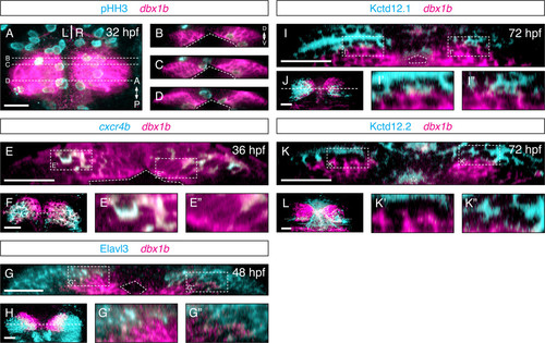
Dbx1b marks a proliferative periventricular domain in the epithalamus. (A) A dorsal view of the epithalamus showed dbx1b and phosphohistone H3 (pHH3) expression. (B-D) Coronal optical sections revealed that dbx1b-positive cells are pHH3-positive. (E) A presumptive habenular precursor marker, cxcr4b, showed partial overlaps with dbx1b. Significantly, the co-expression domain (E′) was more dorsolateral while the dbx1b-only domain (E′′) was along the ventricle. (F) A dorsal view of dbx1b and cxcr4b co-expression. (G-G′′)dbx1b expression showed very little overlap with the neuronal marker Elav3l. (H) A dorsal view of dbx1b- and Elav3l-expressing domains. (I-I′′ and K-K′′) No overlapped expression was observed between dbx1b and markers of differentiated habenular neurons, Kctd12.1 and Kctd12.2. (J and L) Dorsal views of dbx1b- and Kctd12.1/12.2-expressing domains. The ventricle is marked by angled dashed lines. Insets are shown with dashed rectangles. Scale bars are 50 μm.
|