- Title
-
Dbx1b defines the dorsal habenular progenitor domain in the zebrafish epithalamus
- Authors
- Dean, B.J., Erdogan, B., Gamse, J.T., Wu, S.Y.
- Source
- Full text @ Neural Dev.
|
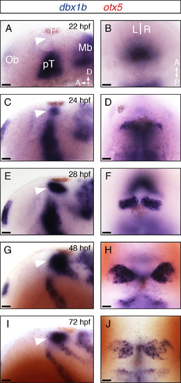
Dbx1b is expressed throughout habenular development. (A-J) Lateral and dorsal views of dbx1b expression (blue) during early brain development. Dorsal diencephalic expression of dbx1b appeared shortly after 22 hpf and continued through 72 hpf (arrowhead). otx5 (red) marks the pineal complex (P). Ob, olfactory bulb; pT, prethalamus; Mb, midbrain. Scale bars are 10 μm. EXPRESSION / LABELING:
|
|
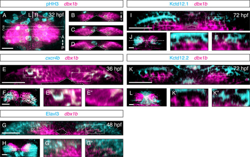
Dbx1b marks a proliferative periventricular domain in the epithalamus. (A) A dorsal view of the epithalamus showed dbx1b and phosphohistone H3 (pHH3) expression. (B-D) Coronal optical sections revealed that dbx1b-positive cells are pHH3-positive. (E) A presumptive habenular precursor marker, cxcr4b, showed partial overlaps with dbx1b. Significantly, the co-expression domain (E′) was more dorsolateral while the dbx1b-only domain (E′′) was along the ventricle. (F) A dorsal view of dbx1b and cxcr4b co-expression. (G-G′′)dbx1b expression showed very little overlap with the neuronal marker Elav3l. (H) A dorsal view of dbx1b- and Elav3l-expressing domains. (I-I′′ and K-K′′) No overlapped expression was observed between dbx1b and markers of differentiated habenular neurons, Kctd12.1 and Kctd12.2. (J and L) Dorsal views of dbx1b- and Kctd12.1/12.2-expressing domains. The ventricle is marked by angled dashed lines. Insets are shown with dashed rectangles. Scale bars are 50 μm. EXPRESSION / LABELING:
|
|
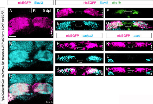
Lineage labeling shows dbx1b-positive cells give rise to only dorsal habenular neurons. (A-C) A dbx1bBAC:cre transgene lineage-labeled (magenta) nearly all Elav3l-positive neurons in the habenulae (cyan). See text for details. The bright area marked with X was due to trapped debris, not actually signal. (D-G) In the same lineage-labeling experiment, an Elav3l-negative domain corresponding the habenular progenitor domain, which is labeled by dbx1b expression (green), was clearly discernible as shown by coronal sections. (H-J) Lineage-labeled cells were restricted within the cadps2-positive dorsal habenular domain except right along the ventricle. (K-M) Lineage-labeled cells were excluded from the aoc1-positive ventral habenulae. Scale bars are 50 μm. EXPRESSION / LABELING:
|
|
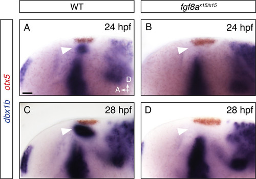
Fgf8a mutants fail to express dbx1b in the epithalamus. (A-D) In situ hybridization for dbx1b (blue) in wildtype and fgf8a mutant embryos. otx5 marks the pineal complex. Scale bars are 10 μm. |
|
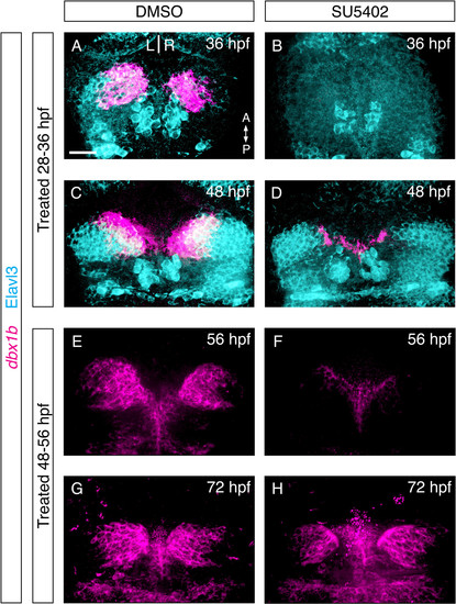
Sustained fibroblast growth factor (FGF) signaling is required for dbx1b expression. (A-D) Eight-hour treatment of embryos with the FGF receptor antagonist SU5402 abolished dbx1b expression, however expression began to return 12 hours after treatment. (E-H) Similar results were seen when FGF receptor blockade was initiated after dbx1b expression began. Generation of Elav3l-positive habenular cells resumed following drug washout at both early and late time points (D, H). Scale bars are 50 μm. EXPRESSION / LABELING:
|
|
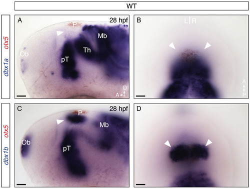
Dbx1b is expressed in the dorsal diencephalon. Lateral and dorsal views of a 28 hpf wildtype embryo. (A and B) In situ hybridization for dbx1a (blue) revealed several expression domains, including the olfactory bulb (Ob), prethalamus (pT), thalamus (Th), and midbrain (Mb) throughout the brain, but no expression in the dorsal diencephalon (arrow heads). (C and D) dbx1b transcript (blue) was expressed in a similar pattern but with greatly reduced expression in thalamus and robust expression in the dorsal diencephalon and olfactory bulb. otx5 (red) marks the pineal complex (P), a component of the dorsal diencephalon. Scale bars are 10 μm. EXPRESSION / LABELING:
|
|
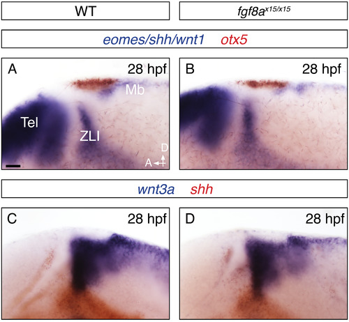
Fgf8a mutants show normal overall brain patterning. (A-B) In fgf8a mutants there were no major anterior-posterior patterning defects observed. eomes, shh and wnt1 mark the telencephalon (Tel), zona limitans intrathalamica (ZLI) and midbrain (Mb) respectively. otx5 marks the pineal complex. (C-D) Dorsal-ventral patterning was also unaffected in fgf8a mutants. wnt3a (blue) marks the ZLI and midbrain and shh (red) marks the ZLI. Scale bars are 10 μm. |

