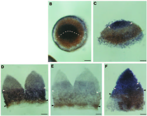
Effect of lithium treatment upon rtk gene expression. (A) Schematic of a severely dorsalized (radialized) embryo at about 12 hours of development. The embryo is symmetrical around its circumference and forms a mound of cells on top of the yolk. The dotted line indicates the extent of the yolk inside the domed embryo. (B-F) Completely radialized embryos labelled with antibody to Ntl (brown) and probes to rtk genes or goosecoid (blue). The approximate anterior limits of Ntl protein and posterior limits of rtk gene expression are indicated with white and black arrowheads respectively. (B) Animal pole view of an embryo labelled with antibody to Ntl plus an RNA probe to goosecoid. Goosecoid is expressed in the most anterior hypoblast at the top of the dome, Ntl protein is present in a ring around the goosecoid-expressing cells. The dotted line indicates the cuts made in such an embryo to produce a flat mount similar to that in C. D-F show similar flat mounts. (C) Ntl plus goosecoid. (D) Ntl plus rtk1. (E) Ntl plus rtk2. (F) Ntl plus rtk3. Scale bars, 50 Ám. Abbreviations: a, anterior; p, posterior; y, yolk
|

