
FIGURE
Fig. S1
Fig. S1
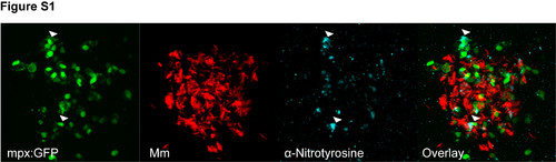
Anti-nitrotyrosine predominantly labels neutrophils in granulomas. Example fluorescent micrographs of anti-nitrotyrosine staining performed on 4 dpi granuloma structures after infection with Mm. The staining colocalized, in the main, with the mpx:GFP fluorescence of neutrophils. Two brightly stained example cells are indicated by the white arrow heads. |
Expression Data
Expression Detail
Antibody Labeling
Phenotype Data
Phenotype Detail
Acknowledgments
This image is the copyrighted work of the attributed author or publisher, and
ZFIN has permission only to display this image to its users.
Additional permissions should be obtained from the applicable author or publisher of the image.
Full text @ PLoS One