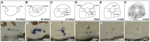
Semicircular canal morphogenesis in the zebrafish ear. (A-F) Sketches of the developing semicircular canal system in the wild-type zebrafish ear, showing projection outgrowth, fusion and pillar formation. (G-L) Expression of vcana in the wild-type ear. (A,G) At 48 hpf, both anterior (A) and posterior (P) projections have begun to grow, and to express vcana at their tips. (B,H) The lateral projection, with its A and P bulges, is present by 50 hpf. The bulges and projections express vcana strongly at this time. The apparent downward growth of the A projection in H may be an artefact of fixation. (C,I) From 57-68 hpf, the A and P projections and bulges fuse to form the A and P pillars. The lateral projection now forms a ventral (V) bulge, and a V projection develops. Expression of vcana is downregulated in the A and P pillars, but is now strongly expressed in the V bulge and projection. (D,J) As fusion is completed at <70 hpf, expression of vcana is downregulated in all pillars. Expression remains in the dorsolateral septum (DLS) at 84 hpf. (E,K) At 72 hpf, all three pillars are fused [timing of fusion was slightly later than previously reported (Waterman and Bell, 1984)]. (F,L) At 4-5 dpf, only a trace of vcana expression remains in the DLS. Grey shading indicates the canal lumens. The positions of the cristae are shown. Abbreviations: A, anterior; DLS, dorsolateral septum; P, posterior; proj., projection; ssc, semicircular canal; V, ventral. Scale bar: 50 μm.
|
