
Fig. S14
- ID
- ZDB-FIG-131010-23
- Publication
- D'Aniello et al., 2013 - Depletion of Retinoic Acid Receptors Initiates a Novel Positive Feedback Mechanism that Promotes Teratogenic Increases in Retinoic Acid
- Other Figures
- All Figure Page
- Back to All Figure Page
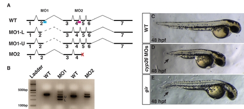
Characterization of cyp26a1 splice-blocking MOs used in experiments. (A) Schematic of the cyp26a1 locus and the intron-exon boundaries targeted by the different cyp26a1 MOs. Blue bar indicates MO1. Red bar indicates MO2. MO1 primarily causes usage of two in-frame cryptic splice sites. Dashed lines indicate the alternate introns cause by the cryptic splices induced from MO1. MO2 causes the introduction of a premature stop codon (red X). (B) RT-PCR for the WT cyp26a1 transcripts and alternate transcripts induced from the different MOs. U and L indicate bands depicted in A. (C) Control sibling embryo. (D) Embryos injected with cocktail of cyp26a1 MO1+2. Co-injection of cyp26a1 MO1 and MO2 causes a phenotype equivalent to or stronger than the cyp26a1/giraffe (gir) mutant (E). Injection of the individual MOs causes the phenotypes consistent with cyp26a1 loss of function at low frequency (data not shown). A suboptimal dose of the cyp26a1 MO cocktail was used for functional interaction experiments with RARαb1 (Figure 4). Arrows in D and E indicate shortened tail. Views in C?E are lateral with anterior right. |