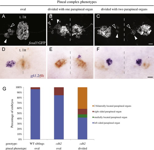
n-cadherin (cdh2vu125) mutants exhibit left-right asymmetry defects. ((A)?(C)) Confocal images of the immunofluorescence labeling of foxd3:GFP positive embryos at 2 dpf. foxd3:GFP is expressed in the pineal and parapineal (white arrowheads) organs. Axonal projections from parapineal neurons are denoted with open arrowheads. ((D)?(F)) Two-color in situ hybridization showing parapineal cells expressing gfi1.2 (blue) and pineal cells expressing flh (red) in 2 dpf embryos. The dashed lines represent the embryonic midline. ((A) and (D)) WT embryos have a fused, oval pineal organ and a single left sided parapineal organ. ((B) and (E)) Approximately half of the cdh2 mutants with a divided pineal organ have only a single parapineal organ. ((C) and (F)) Approximately half of the cdh2 mutants with a divided pineal organ have two bilaterally located parapineal organs. All images are dorsal views with representative images shown. All scale bars=20 μm. (G) Graph of parapineal placement in WT siblings (n=261), cdh2 mutants with oval shaped pineal (n=46) and cdh2 mutants with divided pineal (n=114).
|
