
Fig. 2
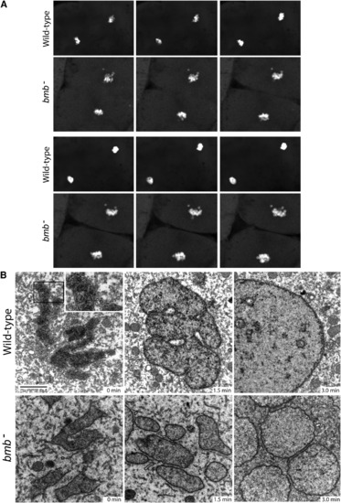
Nuclear Membrane Fusion Is Disrupted in bmb Mutants(A) Frames from time-lapse experiments at the telophase-interphase transition demonstrate that chromatin bodies normally coalesce in WT (top) but fail to do so in bmb (bottom). Note: six frames were selected (from Movies S1 and S2) to best align the sequence of events between WT and bmb at the telophase-interphase transition.(B) Electron microscopy of WT versus bmb at the telophase-interphase transition. Embryos at the 128-cell stage were fixed at 90 s intervals for transmission electron microscopy (TEM). Black bar, 2 microns. The inset in WT (0 min) is enlarged 2� to show the double-membrane nuclear envelope. For each time point, n = 2 embryos. Multiple cells from each embryo were examined in both WT and bmb. |
| Fish: | |
|---|---|
| Observed In: | |
| Stage: | 32-cell |
Reprinted from Cell, 150(3), Abrams, E.W., Zhang, H., Marlow, F.L., Kapp, L., Lu, S., and Mullins, M.C., Dynamic Assembly of Brambleberry Mediates Nuclear Envelope Fusion during Early Development, 521-532, Copyright (2012) with permission from Elsevier. Full text @ Cell