
Fig. 4
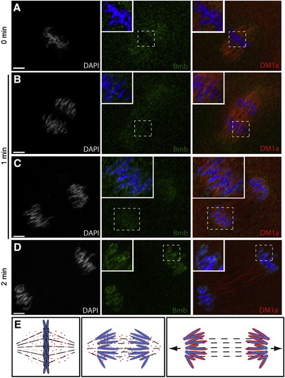
Bmb Protein Dynamics during Mitosis(A) Bmb protein localizes as distinct foci to the mitotic spindle region during metaphase (n = 4).(B) During anaphase, Bmb is interspersed between the separating chromosomes (inset; n = 3) and in the region of the mitotic spindle.(C) Later in anaphase, Bmb foci are decorating the chromosomes (inset; n = 3).(D) At the 2 min time point, Bmb surrounds the individual chromosomes, which are still separating (n = 5).(E) Schematic summarizing Bmb localization during metaphase to anaphase/telophase. Arrows in the right panel indicate that the karyomeres will continue to move to their final central position in the cell. 0 min corresponds to metaphase at the 32- to 64-cell transition. Bmb (green), microtubules (DM1a, red), and chromosomes (DAPI, white and blue in merge).Scale bar, 5 μm, and insets show 2� enlargements. See also Figure S2. |
| Gene: | |
|---|---|
| Antibody: | |
| Fish: | |
| Anatomical Term: | |
| Stage: | 32-cell |
Reprinted from Cell, 150(3), Abrams, E.W., Zhang, H., Marlow, F.L., Kapp, L., Lu, S., and Mullins, M.C., Dynamic Assembly of Brambleberry Mediates Nuclear Envelope Fusion during Early Development, 521-532, Copyright (2012) with permission from Elsevier. Full text @ Cell