|
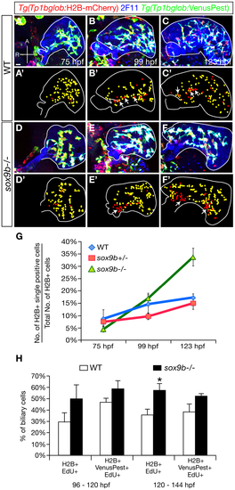
The pattern of Notch signaling activity is altered in sox9b mutants. (A?F) Expression of Tg(Tp1bglob:H2B-mCherry) (red), 2F11 (blue), and Tg(Tp1bglob:VenusPest) (green) in wild-type (A?C) and sox9b mutant (D?F) larvae at 75 (A, D), 99 (B, E), and 123 hpf (C, F). At each time point, 6 larvae of each genotype were analyzed. (A2?F2) Diagrams showing the distribution of Tg(Tp1bglob:H2B-mCherry);Tg(Tp1bglob:VenusPest)-double positive cells (yellow) and Tg(Tp1bglob:H2B-mCherry)-single positive cells (red) in (A?F). Livers are outlined by solid white line. In wild-type livers, expression of Tg(Tp1bglob:H2B-mCherry) and Tg(Tp1bglob:VenusPest) largely overlaps at 75 hpf (A, A′). At 99 and 123 hpf (B?B′, C?C′), a few Tg(Tp1bglob:H2B-mCherry)-single positive cells (arrows) appear along the intrahepatic duct that connects to the extrahepatic system. In sox9b mutants, Tg(Tp1bglob:H2B-mCherry);Tg(Tp1bglob:VenusPest)-double positive cells and Tg(Tp1bglob:H2B-mCherry)-single positive cells show similar distribution as in wild-type at 75 and 99 hpf (D?D′, E?E′). However, at 123 hpf (F, F′), we observed big clusters of Tg(Tp1bglob:H2B-mCherry)-single positive cells in the mutant livers (arrows). (G) Percentages (averageħSEM) of Tg(Tp1bglob:H2B-mCherry)-single positive cells relative to the total number of Tg(Tp1bglob:H2B-mCherry)-expressing cells. Whereas wild-type and sox9b heterozygous livers contained similar percentages of Tg(Tp1bglob:H2B-mCherry)-single positive cells at all stages examined (p>0.4), this percentage was significantly higher in sox9b mutants at 123 hpf (p<0.0005). (H) Percentages (averageħSEM) of Tg(Tp1bglob:H2B-mCherry)-single positive cells or Tg(Tp1bglob:H2B-mCherry);Tg(Tp1bglob:VenusPest)-double positive cells that were labeled by EdU. EdU incubation was conducted from 96 to 120 hpf or from 120 to 148 hpf. Under both conditions, the hepatic Notch responsive cells in sox9b mutants showed higher EdU incorporation compared to wild-type, and the difference was more pronounced for Tg(Tp1bglob:H2B-mCherry)-single positive cells. 7 wild-types and 7 sox9b mutants were examined for each experimental condition. Asterisks indicate statistical significance: *, p<0.05. (A?F) All images are projections of confocal z-stacks. Ventral views, anterior (A) to the top. Scale bar, 20 μm.
|

