
Fig. S2
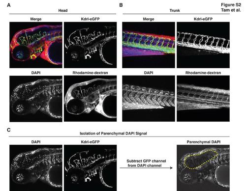
Zebrafish Have a Functional Blood-Brain Barrier at 3 Days Post-Fertilization, Related to Figure 2 (A-B) Upon common cardinal vein microinjection of DAPI (350 Da) and rhodamine-dextran (10 kDa) into Tg(kdrl:egfp) embryos, trunk peripheral tissue leakage of these tracers is widespread whereas within the brain, tracers are tightly restricted within blood vessels. For instance, rhodamine-dextran remains localized within the lumen of brain vasculature and DAPI labels only vascular nuclei but not parenchymal nuclei within the brain. Results representative of at least three independent experiments are shown. Endothelial cells are green, DAPI is blue, rhodamine-dextran is red. (C) Methodology for subtraction blending. DAPI positive brain parenchymal nuclei were visually isolated using Adobe Photoshop CS5 subtraction blending whereby DAPI+GFP+ double-positive signal (DAPI labeling of brain vasculature) was computationally removed from the DAPI channel. |
Reprinted from Developmental Cell, 22(2), Tam, S.J., Richmond, D.L., Kaminker, J.S., Modrusan, Z., Martin-McNulty, B., Cao, T.C., Weimer, R.M., Carano, R.A., van Bruggen, N., and Watts, R.J., Death Receptors DR6 and TROY Regulate Brain Vascular Development, 403-417, Copyright (2012) with permission from Elsevier. Full text @ Dev. Cell