
Fig. 4
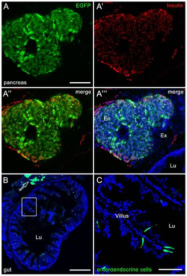
Section from Tg(nrd:egfp)/albino zebrafish showing nrd:egfp trangene expression in the endocrine pancreas (A?A′ ′′) and gut (B?C). (A?A′ ′′). Immunolocalization of EGFP (A, green) co-labels with Insulin (A′, red) in the endocrine pancreas (A′ ′′, En). Co-labeling with TO-PRO-3 shows all nuclei (A′ ′′, blue) and indicates the surrounding exocrine pancreas (A′ ′′, Ex) and adjacent lumen of the gut (A′ ′′, Lu). (B) EGFP expression can be visualized in enteroendocrine cells within each villus and in the surrounding smooth muscle. The adjacent pancreas is also visible (arrow). (C) High magnification image of the box in panel B. |