
FIGURE
Fig. 3
Fig. 3
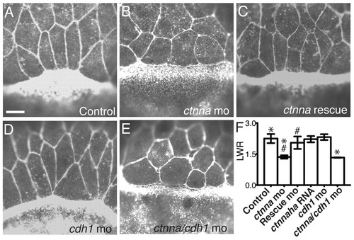
αE-catenin depletion causes defects in EVL cell morphology. (A-E) Confocal images of cells at EVL margin at 80% epiboly stained for F-actin with rhodamine phalloidin. Scale bar: 10 μm. (F) Quantification of length-width ratio (LWR) of cells at the EVL margin. Three independent experiments, n=40 cells from eight embryos. *P<0.002; #P<0.003. Error bars indicate s.d. |
Expression Data
Expression Detail
Antibody Labeling
Phenotype Data
| Fish: | |
|---|---|
| Knockdown Reagents: | |
| Observed In: | |
| Stage: | 75%-epiboly |
Phenotype Detail
Acknowledgments
This image is the copyrighted work of the attributed author or publisher, and
ZFIN has permission only to display this image to its users.
Additional permissions should be obtained from the applicable author or publisher of the image.
Full text @ Development