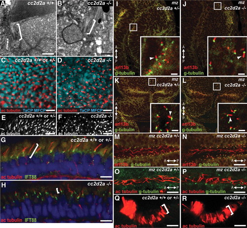
cc2d2a is not required for ciliogenesis. (A and B) Connecting cilia (brackets) in wild-type (A) and cc2d2a-/- (B) photoreceptors demonstrated by transmission electron microscopy. (C and D) Apical view of ciliary axonemes labeled with acetylated tubulin antibodies (red) in wild-type (C) and cc2d2a-/- (D) whole-mount eyes in which cone outer segments are labeled with tg(TaCP:MCFP). (E and F) Peripheral views of the same eyes as in (C and D) showing only the acetylated tubulin staining. Note the shorter axonemes in cc2d2a-/- eyes (F) compared with wild-type (E). (G and H) Ciliary axonemes labeled with acetylated tubulin (red) and Ift88 (green) antibodies in wild-type (G) and cc2d2a-/- (H) retinal cryosections. Brackets highlight axonemal length. Embryos are 5 d.p.f. in (A?F) and 7 d.p.f. in (G and H). (I?N) Immunofluorescence with Arl13b antibody (red) highlighting cilia and with gamma-tubulin antibody (green) highlighting basal bodies in 24 h.p.f. whole-mount mz cc2d2a+/- (I, K and M) and mz cc2d2a-/- (J, L and N) embryos showing retinal neuronal progenitor cilia (arrowheads in insets of I and J), cerebellar neuronal progenitors (arrowheads in insets of K and L) and floorplate cilia (M and N). (O and P) Pronephric duct cilia stained with acetylated tubulin (red) and gamma-tubulin (green) antibodies in 48 h.p.f. whole-mount mz cc2d2a+/- (O) and mz cc2d2a-/- (P) embryos. The anterior?posterior axis is indicated by the arrows. (Q and R) Acetylated tubulin antibody staining of olfactory pit cilia at 3 d.p.f. in whole-mount wild-type (Q) and cc2d2a-/- (R) embryos. (I?L) and (Q and R) Single-confocal sections. (C?F) and (M?P) Projected stacks of confocal sections. Scale bars are 500 nm in (A and B), 5 �m in (C and D), 10 �m in (E and F), 4 �m in (G and H), 2 �m in (I?L), 10 �m in (M and N) and 5 �m in (O?R).
|
