
Fig. 5
- ID
- ZDB-FIG-111117-17
- Publication
- Xi et al., 2011 - Transgenic zebrafish expressing green fluorescent protein in dopaminergic neurons of the ventral diencephalon
- Other Figures
- All Figure Page
- Back to All Figure Page
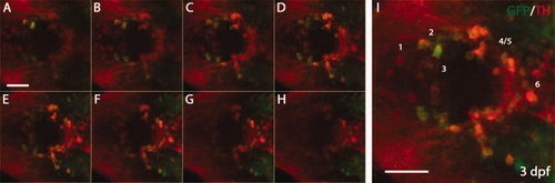
Whole-mount immunostaining for green fluorescent protein (GFP) and tyrosine hydroxylase (TH) in 3 days postfertilization (dpf) Tg(dat:EGFP) larvae. The 3 dpf Tg(dat:EGFP) larvae were stained with anti-GFP and anti-TH antibodies, followed by confocal microscopy. GFP-positive cells and TH-positive cells are shown in green and red respectively. A?H: A series of confocal images focusing at different levels in the ventral diencephalon (vDC). I: Projection of confocal images A?H. Numbers in I indicate different DA neuron groups (1?6) in vDC. The animals are shown as dorsal views with anterior to the left. Scale bars = 25 μm. |
| Genes: | |
|---|---|
| Antibody: | |
| Fish: | |
| Anatomical Terms: | |
| Stage: | Protruding-mouth |