
|
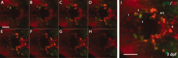
Fig. 5
Whole-mount immunostaining for green fluorescent protein (GFP) and tyrosine hydroxylase (TH) in 3 days postfertilization (dpf) Tg(dat:EGFP) larvae. The 3 dpf Tg(dat:EGFP) larvae were stained with anti-GFP and anti-TH antibodies, followed by confocal microscopy. GFP-positive cells and TH-positive cells are shown in green and red respectively. A–H: A series of confocal images focusing at different levels in the ventral diencephalon (vDC). I: Projection of confocal images A–H. Numbers in I indicate different DA neuron groups (1–6) in vDC. The animals are shown as dorsal views with anterior to the left. Scale bars = 25 μm.