
Fig. 4
- ID
- ZDB-FIG-111117-16
- Publication
- Xi et al., 2011 - Transgenic zebrafish expressing green fluorescent protein in dopaminergic neurons of the ventral diencephalon
- Other Figures
- All Figure Page
- Back to All Figure Page
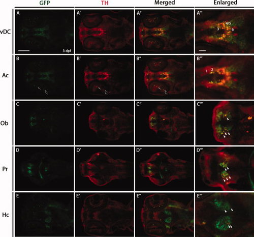
Double immunostaining for green fluorescent protein (GFP) and tyrosine hydroxylase (TH) in 3 days post-fertilization (dpf) Tg(dat:EGFP) larvae. Horizontal cryosections of 3 dpf Tg(dat:EGFP) larvae were stained with anti-GFP and anti-TH antibodies. GFP-positive cells, TH-positive cells and GFP/TH-positive cells are shown in green, red and yellow, respectively. The following abbreviations are used: olfactory bulb (Ob), pretectum (Pr), ventral diencephalon (vDC), amacrine cells (Ac) and caudal hypothalamus (Hc). Numbers in A23 and B23 indicate different groups (1–6) of DA neurons in the vDC. Arrows show GFP/TH-positive cells in the retina. Arrowheads show GFP/TH-positive cells in the Ob, Pr and Hc. Section (A–A23) and section (B–B23) are the same section, focusing on different cell groups when imaging. All sections are shown as anterior to the left. Scale bars = 100 μm in A–A3,B–B3,C–C3,D–D3,E–E3 25 μm in A23,B23,C23,D23,E23. |
| Genes: | |
|---|---|
| Antibody: | |
| Fish: | |
| Anatomical Terms: | |
| Stage: | Protruding-mouth |