
Fig. 6
- ID
- ZDB-FIG-110811-17
- Publication
- Wright et al., 2011 - DeltaC and DeltaD interact as Notch ligands in the zebrafish segmentation clock
- Other Figures
- All Figure Page
- Back to All Figure Page
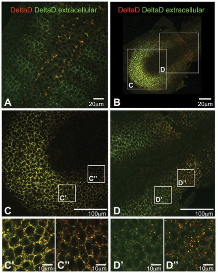
Optical sections of flat-mounted embryos stained to determine the ratio of extracellular to intracellular DeltaD. zdd2 staining before permeabilisation is in green and that after permeabilisation is in red. A low red:green ratio indicates extracellular staining, whereas a high red:green ratio indicates intracellular. (A) Trunk region of a 16 hpf wild-type zebrafish embryo: DeltaD is mainly intracellular and granular in presumptive neuroblasts of the neural tube, but is mainly at the cell surface in the anterior part of each somite. (B-D′′) PSM of a 10-somite stage wild-type embryo. In the posterior PSM, DeltaD is mainly located at the cell membrane (yellow cell outlines in C-C′′); in the anterior PSM DeltaD is predominantly intracellular (red dots in D-D′′). |