
FIGURE
Fig. 5
- ID
- ZDB-FIG-110811-16
- Publication
- Wright et al., 2011 - DeltaC and DeltaD interact as Notch ligands in the zebrafish segmentation clock
- Other Figures
- All Figure Page
- Back to All Figure Page
Fig. 5
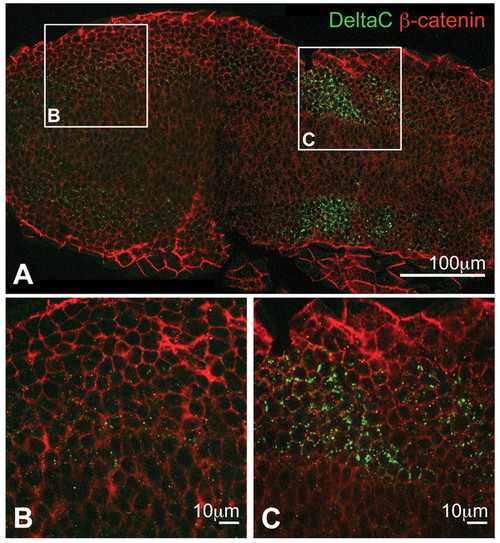
Optical sections of PSM immunostained for DeltaC and β-catenin in a flat-mounted, 10-somite stage wild-type zebrafish embryo. (A) Low-magnification overview. DeltaC, green; β-catenin, red. (B,C) Higher magnifications of the boxed regions in A showing posterior (B) and anterior (C) presomitic mesoderm (PSM). DeltaC staining is always observed as intracellular puncta, with very faint expression in the posterior but strong upregulation in the anterior PSM. |
Expression Data
Expression Detail
Antibody Labeling
Phenotype Data
Phenotype Detail
Acknowledgments
This image is the copyrighted work of the attributed author or publisher, and
ZFIN has permission only to display this image to its users.
Additional permissions should be obtained from the applicable author or publisher of the image.
Full text @ Development