
Fig. S2
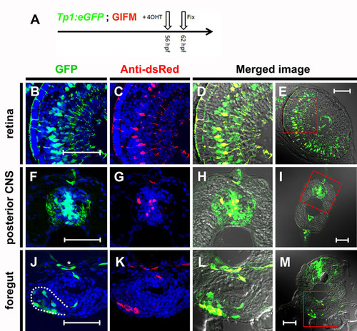
Tp1 Cre-driver restricts recombination in 56 hpf embryos to Notch-responsive cells. (A) As schematized, 4OHT was added at 56 hpf and the embryos were fixed 6 hours later (62 hpf). Again, lineage tracer was detected by immunofluorescence using an antibody to dsRed. (B-M) transverse sections through 4OHT-treated 62 hpf GIFM/Tp1:eGFP embryo. Images are rendered confocal micrographs. Scale bars: 50 μm. (B,F,J) Expression from the Tp1:eGFP transgene marks Notch-responsive cells in green. (C,G,K) Immunofluorescent staining for Hmgb1-mCherry marks lineage traced cells in red. Owing to DAPI staining, all nuclei are marked in blue. (D,H,L) Red and green images merged with bright-field images show all lineage-traced cells are also still Notch responsive. (E,I,M) Lower magnification images show orientation for views in B-D,F-H,J-L respectively. (B-E) Image of a section through the retina where Notch activity marks Müller glia cells. (F-I) Section through trunk of an embryo with Notch activity in cells of the developing central nervous system (CNS). (J-M) Images of a section through the trunk of the embryo at the level of the pancreas. The pancreas is outlined by a broken white line in J and the position of the dorsal aorta is highlighted by an asterisk. |