
Fig. S1
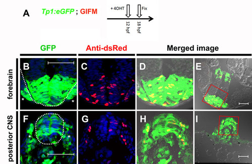
Tp1 Cre-driver restricts recombination in 12 hpf embryos to the Notch-responsive cells. (A) To determine the fidelity of Cre expression in Tp1:creERT2 at 12 hpf, embryos were generated that were transgenic for the GIFM components, as well as for the Notch-responsive marker Tp1:eGFP. As many cells are only transiently Notch responsive, it was necessary to observe co-expression of both eGFP marker and lineage tracer as soon as possible after the addition of 4OHT. As schematized, 4OHT was added at 12 hpf and the embryos were fixed 6 hours later (18 hpf). Six hours is too short a period of time to be able to detect the lineage tracer directly by fluorescence. Instead Hmgb1-mCherry was detected by immunofluorescence using an antibody to dsRed. (B-I) Transverse sections through 4OHT-treated 18 hpf GIFM; Tp1:eGFP embryo. Images are rendered confocal micrographs. Scale bars: 50 μm. (B,F) Expression from the Tp1:eGFP transgene marks Notch-responsive cells in green. (C,G) Immunofluorescent staining for Hmgb1-mCherry marks lineage traced cells in red. Owing to DAPI staining, all nuclei are marked in blue. (D,H) Red and green images merged with bright-field images show all lineage-traced cells are also still Notch-responsive. (E,I) Lower magnification images show orientation for views in B-D,F-H, respectively. (B-E) Section through developing forebrain with the neural epithelium of the telencephalon highlighted with broken white line in B with sensory placodes marked (*). (F-I) Section through posterior of embryo with developing spinal cord of the central nervous system (CNS) highlighted with a broken white line in F and pre-somitic mesoderm marked (*). |