|
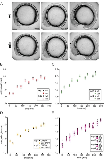
Axial elongation is not changed in Delta-Notch mutant and DAPT-treated embryos
(A) Stills from time-lapse movies [8, 9] of wildtype and mib embryos. White lines: line along which axial length was measured. Arrow: bulge in mutant brain due to neuronal hyperplasia. Bracket: region containing irregular somite boundaries in the mutant. Lateral view with anterior to the left. Scale bar = 100 μm. (B-E) Plots of embryo length (mean ± 95% confidence interval, CI) vs. elapsed recording time for different experimental conditions, n ≥ 4 embryos for each data point. The time points at which wildtype embryos reached the 4 or 10 somite stage are indicated. Values for control and mutant or DAPT-treated embryos were not significantly different (p > 0.01 in all cases as assessed by Student?s t-test) at any time point or for any of the experimental conditions.
|