FIGURE

Fig. S1

ID
ZDB-FIG-100806-84
Publication
Herrgen et al., 2010 - Intercellular Coupling Regulates the Period of the Segmentation Clock
Other Figures
All Figure Page
Back to All Figure Page
Fig. S1

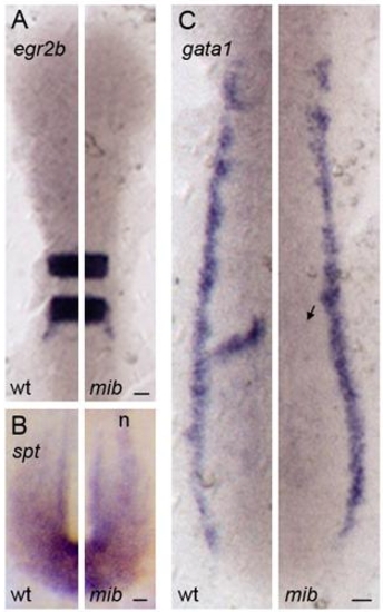
Expression of mesoderm and ectoderm differentiation markers in mib embryos
In situ hybridization with ectoderm (A) and mesoderm (B, C) markers in the PSM of wildtype/heterozygous and mib embryos at the 10 somite stage. Elevated expression of isl1 in (B) in interneurons and Rohon-Beard neurons (n, out of focal plane) and reduced expression of mespa (arrow in C) is observed in mib embryos, either of which was used to distinguish wildtype/heterozygous from mib embryos. Dorsal view with anterior to the top. Expression patterns were assessed in at least five embryos per experimental condition. Scale bars = 25 μm.

Expression Data

Expression Detail
Antibody Labeling
Phenotype Data

Phenotype Detail
Acknowledgments
This image is the copyrighted work of the attributed author or publisher, and ZFIN has permission only to display this image to its users. Additional permissions should be obtained from the applicable author or publisher of the image. Full text @ Curr. Biol.