Fig. 1
- ID
- ZDB-FIG-100525-30
- Publication
- Okuda et al., 2010 - B1 SOX coordinate cell specification with patterning and morphogenesis in the early zebrafish embryo
- Other Figures
- All Figure Page
- Back to All Figure Page
|
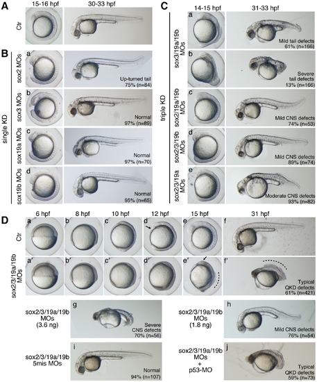
Single, triple, and quadruple knockdowns of sox2/3/19a/19b. Bright-field images of live embryos observed at the indicated time points. All are lateral views. (A) Uninjected control (Ctr) embryos. (B) Single knockdowns of sox2/3/19a/19b. A 1:1 mixture of two MOs (0.9 ng each) targeting one of the four B1 sox genes was injected for a single KD. The percentage of embryos in the same morphological class is indicated in each panel. (C) Triple knockdowns of sox2/3/19a/19b. A mixture of indicated combinations of MOs (i.e., a mixture of six MOs, 5.4 ng in total) was used to simultaneously knockdown three out of the four B1 sox genes. The major classes of morphological defects are shown with the percentage of occurrence. The remaining embryos showed either milder or more severe defects. (D) Uninjected control embryos (a?f) and sox2/3/19a/19b quadruple knockdown (QKD) embryos injected with a mixture of MOs targeting the four B1 sox genes (i.e., a mixture of eight MOs, 7.2 ng in total) (a′?f′). The QKD caused very severe developmental abnormalities: a delay in epiboly, a shortened anterior-posterior axis, and impairment of CNS development (61%). The remaining embryos showed either milder defects (7%), more severe defects (24%) or lethality (8%). The aberrant movement of the anterior prechordal plate (arrows in d and e′) suggests a decreased adhesion of ectodermal cells as well as defects in convergence and extension movements in the QKD embryos. The broken lines (e′ and f′) indicate the dorsal trunk regions where cell dissociation was observed. (g, h) Dose-dependent effects of the MOs used for QKD were examined by injecting reduced amounts of the mixture of MOs targeting the four B1 sox genes (3.6 ng in total [g] and 1.8 ng in total [h]). (i) As a negative control, a mixture of 5-base-mismatch control MOs (i.e., a mixture of eight 5mis-MOs, 7.2 ng in total) was injected. (j) The coinjection of a p53-MO (2 ng) had no impact on the neural defects in the QKD embryos. |

