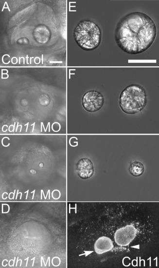
Otolith morphology in Cdh11 knockdown embryos. Zebrafish were injected with control morpholino oligonucleotide (A, E) or cdh11 antisense morpholino oligonucleotide (B-D, F, G), and these embryos (7 dpf) (A-D) and their dissected otolith crystals (E-G) were imaged by DIC microscopy (Nikon diaphot 200: A-D, Plan 20x 0.4 NA objective; E-G, 40x 0.55 NA objective). The otoliths shown in E-G were the same otoliths seen in the inner ears in A-C, respectively. Live embryos mounted in embryo medium (A-D) and otoliths were mounted in PBS. Note that the crystal diffraction patterns are similar, comparing control and morphant otoliths, but the sizes of morphant otolith crystals are reduced (F, G). The more caudal otolith (associated with the posterior macula) was smaller than the more rostral otolith in some morphants (for example, in C and G). Cdh11 immunofluorescence of 3-dpf wholemount-labeled embryos mounted in PBS (H) shows annular patterns in the growing otolith (arrow), and abundant vesicular structures, many associated with the surface of the growing otolith (note: speckle pattern on otolith surface). Two photon microscopy using a BioRad MRC1024, 60x W 1.2 NA, 800-nm illumination, pixel size 0.39 μm, step size 0.4 μm. A projection image of 50 optical sections was made using Voxx Software in alpha blending mode. Arrowhead highlights a linear array of punctate Cdh11 immunolabeling that may represent association with a kinocilium. Scale bar in A = 50 μm for A-D, and the scale bar in E = 50 μm for E-G. For A-D and H, rostral is left, and dorsal is up. Figure was made with Photoshop 6, applying contrast stretch and unsharp mask.
|

