
Fig. 2
- ID
- ZDB-FIG-090401-2
- Publication
- Sonawane et al., 2009 - Lgl2 and E-cadherin act antagonistically to regulate hemidesmosome formation during epidermal development in zebrafish
- Other Figures
- All Figure Page
- Back to All Figure Page
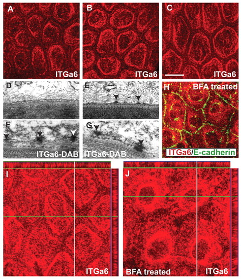
Dynamics of hemidesmosome formation in the zebrafish epidermis. (A-C) Immunostaining using anti-Itga6 antibody in wild-type larvae analysed at 3, 4 and 5 dpf in whole-mount. The localisation of Itga6 to the basal domain increases, while at the lateral domain it progressively decreases during 3-5 dpf. (D-G) Electron microscopy (D,E) and immunoelectron microscopy using anti-Itga6 antibody and nickel-enhanced DAB (F,G) in wild type. Electron-dense hemidesmosomes (arrowheads) are absent from the epidermis at 3 dpf (D) but present at 4 dpf (E). Itga6 localises to intermediate filaments (arrowheads) at 3.5-dpf (F). At 4 dpf, Itga6 becomes incorporated in hemidesmosomes (G). (H) Co-immunostaining using anti-Itga6 (red) and E-cadherin (green) antibodies. (I,J) Immunostaining using anti-Itga6 antibody followed by analysis in x-y and x-z planes. The basally localised Itga6 (I) accumulates around the nucleus after BFA treatment (H,J). Scale bar: 13.5 μm in A-C,H; 200 nm in D-G. |