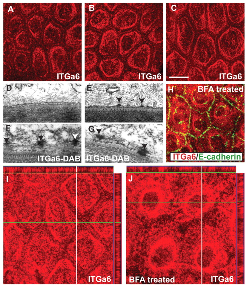
Fig. 2 Dynamics of hemidesmosome formation in the zebrafish epidermis. (A-C) Immunostaining using anti-Itga6 antibody in wild-type larvae analysed at 3, 4 and 5 dpf in whole-mount. The localisation of Itga6 to the basal domain increases, while at the lateral domain it progressively decreases during 3-5 dpf. (D-G) Electron microscopy (D,E) and immunoelectron microscopy using anti-Itga6 antibody and nickel-enhanced DAB (F,G) in wild type. Electron-dense hemidesmosomes (arrowheads) are absent from the epidermis at 3 dpf (D) but present at 4 dpf (E). Itga6 localises to intermediate filaments (arrowheads) at 3.5-dpf (F). At 4 dpf, Itga6 becomes incorporated in hemidesmosomes (G). (H) Co-immunostaining using anti-Itga6 (red) and E-cadherin (green) antibodies. (I,J) Immunostaining using anti-Itga6 antibody followed by analysis in x-y and x-z planes. The basally localised Itga6 (I) accumulates around the nucleus after BFA treatment (H,J). Scale bar: 13.5 μm in A-C,H; 200 nm in D-G.
Image
Figure Caption
Acknowledgments
This image is the copyrighted work of the attributed author or publisher, and
ZFIN has permission only to display this image to its users.
Additional permissions should be obtained from the applicable author or publisher of the image.
Full text @ Development

