Fig. 6
- ID
- ZDB-FIG-090401-7
- Publication
- Sonawane et al., 2009 - Lgl2 and E-cadherin act antagonistically to regulate hemidesmosome formation during epidermal development in zebrafish
- Other Figures
- All Figure Page
- Back to All Figure Page
|
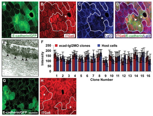
The reduction of Lgl2 levels in ecadMO clones rescues the hemidesmosomal phenotype. (A-D) Co-immunostaining using anti-E-cadherin/GFP (A), anti-Itga6 (B) and anti-Lgl2 (C) antibodies and overlay (D). In ecadMO clones (A), Itga6 does not show enhanced localisation at 6 dpf (B) when Lgl2 levels (C) are reduced by co-injecting lgl2 morpholino. Note that Lgl2 is not completely absent after injection of suboptimal concentrations (100 μM) of lgl2 morpholino (arrowheads in D). Quantification of signal intensities reveals that there are no significant differences in the Itga6 intensities in clonal cells and surrounding host cells (D). Asterisks, epidermal clones. In A and E, the cytoplasmic green fluorescence represents GFP. Our fixation conditions do not lead to complete quenching of GFP fluorescence. (E) Electron micrograph of lgl2-ecadMO clone. (F) The signal intensity for Itga6 was quantified from three host cells and from three cells per clone that received e-cadherin as well as lgl2 (at suboptimal concentrations) morpholinos. The mean intensity for each cell and the standard error are shown. (G,H) Co-immunostaining using anti-E-cadherin (G) and anti-Itga6 (H). This clonal analysis in 3.5-day-old larvae reveals that ecadMO clones (G) do not exhibit enhanced localisation of Itga6 at the basal or lateral domain (H). Scale bar: 27 μm in A-D,G,H; 270 nm in E. |

