
Fig. 3
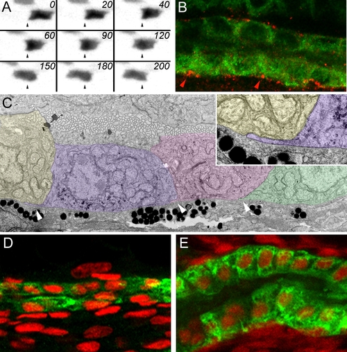
Pronephric Epithelial Cell Migration (A) Time-lapse images of an individual pronephric multiciliated cell in a 2.5 dpf CD41:GFP transgenic fish at 20-min intervals. Dynamic transient cytoplasmic projections are seen from the basal cell surface, most visible in frames 0 min, 60 min, and 120 min. (B) Double immunofluorescence staining of the pronephros in 3-dpf fish with anti-phospho-FAK antibody (red) and anti-alpha6 NaK ATPase (green). Phospho-FAK staining is positive at the epithelial cell interfaces along the basement membrane. (C) Transmission electron microscope images show that migrating epithelial cells send cryptic lamellipodia along the basement membrane in the direction of migration (arrowheads). Here three migrating cells are false colored to distinguish individual cells. Inset shows a single basal cytoplasmic projection. (D and E) The change in epithelial morphology of the proximal kidney (ET33-D10 GFP segment) between 36 hpf (D) and 96 hpf (E). At 36 hpf, the proximal epithelial cells are low cuboidal and the tubular diameter is small. At 96 hpf, the proximal tubule has much larger diameter and the epithelial cells become columnar. The ET33-D10 transgenic embryos were stained with anti-GFP antibody (green Alexa 488 secondary) and DAPI (red pseudocolor). |
| Gene: | |
|---|---|
| Fish: | |
| Anatomical Term: | |
| Stage: | Pec-fin |