
FIGURE
Fig. 5
Fig. 5
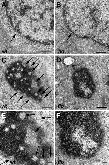
Nuclear ultrastructure in flo mutants. Transmission electron micrographs of nuclei from representative 5 dpf wild type and flo intestinal epithelial cells. (A,B) Intact nuclear envelope in wild type (A) and flo (B). (C?F) Tangential sections through the nuclear envelope showing abundant nuclear pores (arrows) in the wild type larva (C,E) but few if any well defined pores in the flo larva (D,F). (E) and (F) are higher magnification views of (C) and (D), respectively. |
Expression Data
Expression Detail
Antibody Labeling
Phenotype Data
| Fish: | |
|---|---|
| Observed In: | |
| Stage: | Day 5 |
Phenotype Detail
Acknowledgments
This image is the copyrighted work of the attributed author or publisher, and
ZFIN has permission only to display this image to its users.
Additional permissions should be obtained from the applicable author or publisher of the image.
Full text @ PLoS Genet.